Department of Kaumarbhritya, Mahatma Gandhi Ayurveda College Hospital and Research Centre, Datta Meghe Institute of Higher Education & Research, Sawangi, Wardha, Maharashtra, India.
Arun Naphe Khatri
Email: arunkhatri301@gmail.com
Received : Oct 01, 2024 Accepted : Nov 04, 2024 Published : Nov 11, 2024 Archived : www.meddiscoveries.org
Infantile hemangiomas are common vascular tumors occurring in infancy and early childhood, with a prevalence of approximately 5-10% among infants. These hemangiomas develop due to an abnormal proliferation of capillaries, which are densely packed and lack open lumens. During infancy, these tumors exhibit a highly proliferative phase but gradually stabilize over time. Most hemangiomas undergo a slow, spontaneous regression and tend to resolve completely by the age of five. The prevalence is approximately three times higher in females than in males. This report presents a case of a 4-month-old female infant who was brought to us with a progressively enlarging swelling on the left side of her face. There were no noticeable birthmarks at the time of birth, but the swelling appeared and increased in size over time. The lesion was prone to bleeding, even with minor trauma. The eventual regression of the lesion supports the diagnosis of an infantile hemangioma.
Keywords: Infantile hemangioma; Zygomatic bone; Β-adrenergic blockers; Propranolol.
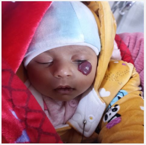
A female child, aged 3 months, was brought to our attention due to swelling and a large area of discoloration on the left side of her face. This condition had developed approximately 1 month after birth and did not appear to affect her functionality. The patient’s growth and development appeared to be on track, and she achieved developmental milestones normally. Upon examination, a dispersed large area of discoloration was observed on the left side of her face, just below the zygomatic bone. The swelling measured 2 x 2 cm in size, with a reddish-brown color and areas of erythema and necrosis in some places (Figure 1). The skin over the tumor had an altered texture and did not appear normal. There were no noticeable oral lesions, and no other significant changes were observed intraorally.
We prescribed propranolol syrup, with a recommended dosage of 1 mg/kg/day for the initial 3 days, followed by an increased dosage of 2 mg/kg/day for the subsequent 10 days.
Differential diagnosis: Vascular malformations, Pyogenic granuloma, Nevus flammeus (Port-Wine Stain), Strawberry nevus (Infantile Hemangioma).
Infantile hemangiomas are benign vascular tumors that typically occur during infancy and early childhood. The term “hemangioma” refers to any vascular tumor-like structure, which can either be present at birth or develop later in life. Hemangiomas can be classified into two main groups. The first group consists of growing lesions that eventually resolve on their own (self-involuting tumors), while the second group comprises malformations characterized by enlarged or abnormal blood vessels that are present at birth and tend to be permanent, not undergoing self-resolution. In neonates, the prevalence of infantile hemangiomas is approximately 2 percent. By the age of one year, the prevalence increases to 10 percent, and in preterm babies weighing less than 1000 grams, it can be as high as 22-30 percent [1,2]. Interestingly, infantile hemangiomas are three times more prevalent in female patients compared to male patients [3,4]. The most common location for infantile hemangiomas in the body is the head and neck, accounting for around 60 percent of cases, followed by approximately 25 percent in the trunk region, and the least commonly in the extremities, which make up around 15 percent of cases. The choice of treatment for hemangiomas depends on the various stages of growth and should be carefully considered in consultation with the child’s parents [5]. The medical management of Infantile Hemangiomas (IH) includes the use of topical corticosteroids, oral medications such as propranolol, corticosteroids, alpha-interferon, anticancer drugs, and imiquimod. Topical corticosteroids can sometimes lead to ulceration, and if the ulceration is deep, significant bleeding may occur. Propranolol and systemic corticosteroids are well-established therapies for treating infantile hemangiomas [6]. Before starting systemic steroid therapy, a thorough pre-treatment evaluation is necessary, including a complete blood count with differential, serum biochemistry, chest X-ray, and urine and stool microscopy to rule out infections, baseline abnormalities, or primary immunodeficiency. Additionally, baseline anthropometric measurements (height and weight) and regular monitoring of blood pressure are crucial throughout treatment [7].
For propranolol therapy, it is important to screen the child for allergies to the medication, as well as conditions such as bronchial asthma, hypoglycemia, hypotension, sinus bradycardia, heart block, and heart failure. Steroid therapy is most effective during the early proliferative phase of hemangioma growth. The mechanism of action of corticosteroids is believed to involve inhibition of vascular endothelial growth factor A (VEGFA) production by proliferating or stem cells in the hemangioma, leading to reduced angiogenesis and tumor growth.
Common side effects of systemic corticosteroid therapy for infantile hemangiomas include growth disturbances, polyuria, loss of appetite, increased susceptibility to infections, excessive hair growth (pilosity), a Cushingoid appearance, oral thrush, gastrointestinal discomfort, and behavioral changes. Oral corcorticosteroids administered at a dose of 2-3 mg/kg/day typically result in a 75% response rate, while doses exceeding 3 mg/kg/day show a higher response rate of 94%, albeit with more severe side effects. Conversely, doses below 2 mg/kg/day tend to yield poor responses and are associated with a rebound phenomenon in about 70% of cases [8,9]. The treatment of hemangiomas must be tailored to the various stages of tumor growth and should be approached with caution, always in consultation with the child’s parents. Ulceration in deeper areas can be particularly painful and problematic. However, most hemangiomas regress naturally over time without intervention, often leaving minimal or no visible marks. In some cases, large hemangiomas may cause visible skin changes due to significant stretching or damage to the skin’s surface, even after regression.
Infantile Hemangiomas (IH) are common benign vascular tumors that occur in infancy and early childhood. In the pediatric population, oral corticosteroids are considered a safe and effective treatment option with minimal serious complications. However, these tumors can cause significant anxiety and discomfort for parents, making proper evaluation and thorough counseling essential in managing both the condition and the concerns of the caregiver.
