1Senior Registrar, Medicine National Hospital, Sri Lanka.
2Consultant Physician, National Hospital of Sri Lanka, Srilanka.
Perera MNSK
Email: nishadiperera19@yahoo.com
Received : Oct 07, 2024 Accepted : Nov 01, 2024 Published : Nov 08, 2024 Archived : www.meddiscoveries.org
Pulmonary veno occlusive disease is a rare cause of pulmonary hypertension which has a relentlessly progressive course with high morbidity and mortality. Because of rarity of the disease, management options are also not well established. We present a case of a 64-year-old lady who came with progressive breathlessness and fatigue; and was found to be having clinical evidence of pulmonary hypertension and right ventricular failure, which was confirmed by echocardiography. Her pulmonary angiogram confirmed pulmonary veno occlusive disease as the cause of pulmonary hypertension while other secondary causes were excluded. She was treated with pulmonary vasodilator therapy cautiously and is currently under follow-up. This patient’s story illustrates pulmonary veno occlusive disease as a rare cause of pulmonary hypertension. It should be considered as a differential diagnosis in patients with pulmonary hypertension where a secondary aetiological factor cannot be found.
Keywords: Pulmonary hypertension; Veno-occlusive disease; Computed tomography pulmonary angiogram.
Progressive breathlessness is a common clinical presentation of patients with pulmonary hypertension. When considering the etiologies for pulmonary hypertension, pulmonary veno occlusive disease is a rare cause categorized under type 1 pulmonary hypertension [1]. Imaging studies and cardiac catheterization helps to confirm the diagnosis [2]. Correct diagnosis is prudent here, as the prognosis is poor and the treatment modalities differ when compared to other causes of pulmonary hypertension this case describes the presentation of a lady with pulmonary hypertension, which turned out to be due to pulmonary veno occlusive disease, and how the diagnosis has been made.
A 64-year-old lady presented with 3 months history of progressive breathlessness. She had diabetes mellitus which was well controlled, while on pre-mixed insulin, without macro vascular or micro vascular complications.
Her symptoms progressed gradually, and upon admission, she was breathless at rest, and further complained of lower limb swelling. Her symptoms didn’t have a diurnal variation, there were no orthopnoea or paroxysmal nocturnal dyspnoea. She noticed an occasional dry cough; but never had fever, sputum or hemoptysis.
Her past medical history was unremarkable for chronic lung diseases; neither had she been exposed to smoking or noxious substances. There was no history of ischemic heart disease, congenital or acquired valvular heart disease, neither there was a history of inflammatory-type joint pains or rashes to suggest an underlying connective tissue disorder. History did not reveal previous events of arterial or venous thrombotic manifestations. Upon admission, the patient had bilateral pitting pedal edema; no clubbing. She was tachypnoeic and, her arterial oxygen saturation was 84% on ambient air. Auscultation revealed vesicular breath sounds bilaterally, symmetrically without added sounds. She had a normal volume pulse at a rate of 80/min. Jugular venous pulsation was elevated. Apical impulse was not displaced and there wasn’t a para-sternal heave. The first heart sound was normal but the pulmonary component of the second heart sound was loud. There were no audible murmurs. The rest of the systemic examination was unremarkable. Urgent arterial blood gas analysis on room air revealed the following findings:
| pH | 7.52(7.35 to 7.45) |
| pCO2 | 41 mmHg (35-45 mmHg) |
| pO2 | 43 mmHg (80-100 mmHg) |
| HCO3 | 33.5 mmol/L(22-16) |
| SpO2 | 84% (95-100%) |
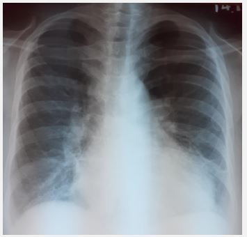
Electrocardiogram (ECG) had; prominent p waves in lead II and aVF, and deep S waves from V1 to V6, which were consistent with pulmonary hypertension. Serial Troponin I values were negative. Chest radiography revealed prominent central pulmonary arteries. There were no interstitial shadows, evidence of consolidation, or effusions (Figure 1).
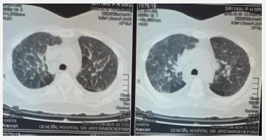
Her full blood count, renal and liver function studies and inflammatory markers were within the normal range. Her autoimmune antibody panel too was negative. Transthoracic echocardiography revealed an ejection fraction of 60%, without regional wall motion abnormalities. The right atrium and ventricle were mildly dilated. There was moderate pulmonary hypertension with a tricuspid regurgitant peak gradient of 40 mmHg. Cardiac valves were normal. Transoesophageal echocardiogram didn’t reveal intra-cardiac shunts. In the technetium perfusion scan, normal homogenous tracer uptake was seen in the upper, mid and lower zones of both lungs. It was concluded that the possibility of chronic thromboembolism is very low. Computed Tomography Pulmonary Angiogram (CTPA) showed dilated right, left and main pulmonary arteries, and reflux of contrast medium into inferior vena cava suggesting pulmonary hypertension. Smooth diffuse interlobular septal thickening with ground glass opacity was noted involving all segments of both lungs. There were no hypodense wedge-shaped areas in lung fields to suggest infractions. It was concluded as pulmonary veno occlusive disease, without any CT features to suggest acute or chronic pulmonary embolism. Her High-Resolution Computed Tomography (HRCT) chest showed interlobular septal thickening with mosaic attenuation, without evidence of interstitial lung disease (Figure 2).
She was managed as pulmonary hypertension due to pulmonary veno occlusive disease, after liaison with the consultant respiratory physician. She was started on sildenafil 25 mg daily dose. The patient improved gradually while on sildenafil; upon discharge from the ward, she was ambulant, breathlessness was only noted upon exertion, pedal edema was settled and her general well-being was improved.
Her arterial blood gas analysis upon discharge is given below:
| pH | 7.44 |
| pCO2 | 36 mmHg |
| pO2 | 59 mmHg |
| HCO3 | 22.7 mmol/L |
| SpO2 | 91.7% |
The patient is currently under follow-up with vasodilator therapy; however, the patient and the family were well-informed of the natural course of the illness while continuing medical care.
This patient who came with progressively worsening breathlessness, was in type 1 respiratory failure on admission without evidence of primary lung pathology. Her cardiovascular examination findings were consistent with pulmonary hypertension with right heart failure. ECG and echocardiogram confirmed pulmonary hypertension. Ischeamic heart disease, intracardiac shunts or valvular lesions were excluded. Normal HRCT excluded primary pulmonary pathologies. CTPA, supported by the normal perfusion scan excluded the probability of pulmonary venous thromboembolism, and it was consistent with the diagnosis of pulmonary veno occlusive disease. Pulmonary veno occlusive disease is a rare cause of pulmonary hypertension [1]. It has a relentlessly progressive course and a poor prognosis than thromboembolic pulmonary hypertension [3]. One-year mortality is around70%, where the mean survival rate falls into 1 to 2 years [4,5].
In pulmonary veno occlusive disease, occlusion of pulmonary venule and capillaries occur by fibrous tissue due to unknown triggers [6]. Intimal thickening involves in venules and small veins at interlobular septae. Pulmonary arterioles will show hypertrophy of the media. Pulmonary lymphatics get dilated [6]. Aetiological factors or autoimmune associations have not being identified still, due to lack of proper case control studies [6,8]. CTPA and HRCT chest are helpful diagnostic modalities which shows characteristic features including diffuse interlobular septal thickening [9]. Histology would definitely confirm the diagnosis, which was not feasible in our clinical context. Right heart catheterization, which could not be performed in this patient, would have confirmed pre capillary pulmonary hypertension with elevated mean pulmonary arterial pressure, elevated pulmonary vascular resistance while pulmonary capillary wedge pressure would be normal [2,3]. Evidence for usage of pulmonary vasodilator therapy for treatment is conflicting because it can cause non cardiogenic pulmonary oedema [3]. Therefore, the recommendation is to use pulmonary vasodilator therapy judiciously, closely monitoring for complications [10]. Our patient tolerated pulmonary vasodilator therapy well, with clinical improvement.
Long term oxygen therapy is indicated in who are hypoxaemic. Lung transplantation is the only proven threapy [11,12].
In conclusion, pulmonary veno occlusive disease remains a rare cause of pulmonary hypertension which is poorly understood yet. Although it is a rare entity, it should be considered as a differential diagnosis in patients with pulmonary hypertension where a secondary aetiological factor cannot be found.
