Clinical Image
Volume 3, Issue 6
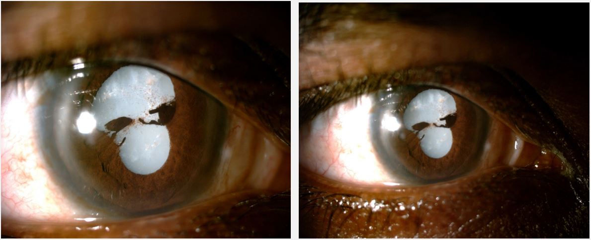
Skull Shape Traumatic White Cataract
Houda Brarou1*; Manal Bouggar1 ; Zakaria Chaabi1 ; Soundouss Sebbata1 ; Lucrece Joannele Eriga Vydalie1 ; Soukaina Laaouina; Samah Sadiki1 ; Nermine Belayachi1 ; Taoufik Abdellaoui2 ; Yassine Mouzari1 ; Abdelbarre Oubaaz1
1Ophtalmology Department, Military Hospital Mohammed-V, Mohammed-V University-Rabat, Morocco.
2Sidi Mohamed Ben Abdellah University, Fes, Morocco.
Corresponding Author :
Houda Brarou
Tel: +21-267-372-1525;
Email: houdaophthalmology@gmail.com
Received : May 18, 2024 Accepted : Jun 15, 2024 Published : Jun 22, 2024 Archived : www.meddiscoveries.org
Citation: Brarou H, Bouggar M, Chaabi Z, Sebbata S, Vydalie LJE, et al. Skull Shape Traumatic White Cataract. Med Discoveries. 2024; 3(6): 1172.
Copyright: © 2024 Brarou H. This is an open access article distributed under the Creative Commons Attribution License, which permits unrestricted use, distribution, and reproduction in any medium, provided the original work is properly cited.
Description
A 32 years old male patient presented to the Ophthalmology Department reporting a progressive decrease in vision in his right eye (OD) in the past year. He reported a history of blunt right eye injury while 18 months ago. At the time of the injury, the patient was asymptomatic. After the incident occurred, the patient did not have a further ophthalmic examination.
The visual acuity of the right eye was reduced to light perception and was 10/10 in the left eye. Intraocular pressures measured in both eyes were within the normal limit. The anterior segment examination of the right eye showed a white dense cataract. After dilation, Iris adhesions to the lens (posterior synechiae) were present at 3 o’clock and 9 o’clock (Figure 1).


