Clinical Image
Volume 3, Issue 6
Blue-Green Inclusions in Neutrophils in a Patient with Pancreatic Necrosis
Tatiyana V Konyuhova; Ekaterina V Trukhina*
Doctor of Clinical and Diagnostic Laboratory of the Dmitry Rogachev National Medical Research Center of Pediatric Hematology, Oncology and Immunology of Ministry of Healthcare of the Russian Federation, Moscow 117997, Russia.
Corresponding Author :
Ekaterina V Trukhina
Tel: +7-977-172-02-50;
Email: Ekaterina-87@bk.ru
Received : May 21, 2024 Accepted : Jun 12, 2024 Published : Jun 19, 2024 Archived : www.meddiscoveries.org
Citation: Konyuhova TV, Trukhina EV. Blue-Green Inclusions in Neutrophils in a Patient with Pancreatic Necrosis. Med Discoveries. 2024; 3(6): 1171.
Copyright: © 2024 Trukhina EV. This is an open access article distributed under the Creative Commons Attribution License, which permits unrestricted use, distribution, and reproduction in any medium, provided the original work is properly cited.
Abstract
Blue-green cytoplasmic inclusions in neutrophils in peripheral blood smears are an important finding described in critically ill patients and associated mainly with acute liver failure and lactic acidosis. Also, blue-green inclusions have been described in patients with multiple organ failure, COVID19 infection, yellow fever and sepsis caused by E. Coli. In this article, we report on a patient who had blue-green inclusions in neutrophils 48 hours before the onset of death.
Keywords: Blue-green inclusions; Death crystals; Lactic acidosis; Neutrophils.
Description
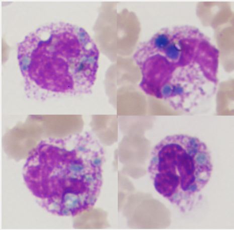
An 11-year-old girl was admitted to the intensive care unit with complaints of muscle weakness, nausea, swelling, and abdominal pain. A history of juvenile dermatopolymiositis, acute course, secondary hemophagocytic syndrome, long-term therapy with glucocorticosteroids, cyclophosphane. According to magnetic resonance imaging of the abdominal cavity, signs of edematous pancreatitis and ascites were revealed. Blood gas readings indicated lactic acidosis (pH 7.10, lactate 11.3 mmol/l), an increase in pancreatic enzymes (pancreatic amylase 84 U/L, lipase 322 U/L, alpha-amylase 123 U/L). A smear of peripheral blood revealed blue-green inclusions in the cytoplasm of neutrophils. Inclusions were often multiple, variable in size and shape (Figure 1). The percentage of neutrophils containing such inclusions was not large and amounted to 1-2%. The patient died 48 hours later. According to the autopsy, the cause of death was pancreatic necrosis.
Blue-green inclusions are an important clinical finding and are found only in blood smears in the cytoplasm of neutrophils, less often monocytes. They were first described by Smith in 1967 [1]. In the literature, this type of inclusions was previously called “death crystals”, since most patients died within 24-48 hours. Modern hypotheses suggest that these inclusions are lipofuscin and appear as a result of phagocytosis of products of hepatocellular damage by neutrophils and monocytes [2]. The identification of these inclusions indicates an extremely serious condition of the patient. Laboratory diagnostic doctors should be aware of their existence and inform the attending physician immediately.
Declarations
Contribution of the authors: Trukhina EV data collection, preparation of a list of references. Konyukhova TV article design development, writing the text of the manuscript.
Conflict of interest: The authors declare no conflict of interest.
Funding: The study was performed without external funding.
References
- Harris VN, Malysz J, Smith MD. Green neutrophilic inclusions in liver disease. J Clin Pathol. 2009; 62(9): 853-4. doi: 10.1136/ jcp.2009.064766. PMID: 19734487.
- Haberichter KL, Crisan D. Green Neutrophilic Inclusions and Acute Hepatic Failure: Clinical Significance and Brief Review of the Literature. Ann Clin Lab Sci. 2017; 47(1): 58-61. PMID: 28249918.


