Short Report
Volume 3, Issue 5
Pediatric Atelectasis: Insights from Diagnostic Imaging
Rebeca Tenajas1; David Miraut2*
1Family Medicine Department, Arroyomolinos Community Health Centre, Arroyomolinos, Spain.
2Advanced Healthcare Technologies Department, GMV Innovating Solutions, Spain.
Corresponding Author :
David Miraut
Email: dmiraut@gmv.com
Received : Mar 15, 2024 Accepted : May 02, 2024 Published : May 09, 2024 Archived : www.meddiscoveries.org
Citation: Tenajas R, Miraut D. Pediatric Atelectasis: Insights from Diagnostic Imaging. Med Discoveries. 2024; 3(5): 1150.
Copyright: © 2024 Miraut D. This is an open access article distributed under the Creative Commons Attribution License, which permits unrestricted use, distribution, and reproduction in any medium, provided the original work is properly cited.
Abstract
This article presents a detailed case study of a 10-year-old child, born in 2013, exhibiting symptoms of a febrile syndrome and subsequent diagnostic findings suggestive of atelectasis through chest radiography and Computed Tomography (CT) scans without intravenous contrast. Atelectasis, a condition characterized by the collapse of lung tissue, represents a significant diagnostic challenge in pediatric patients. This study delves into the radiographic and CT findings, emphasizing the importance of imaging in diagnosing and managing atelectasis.
Keywords: Pediatric atelectasis; Diagnostic imaging; Chest radiography; Lung diseases.
Introduction
Atelectasis, the partial or complete collapse of a part or the entire lung, is a condition often encountered in pediatric medicine. It can arise from various etiologies, including obstruction, infection, or trauma, leading to a reduction in gas exchange and potentially complicating the clinical course of young patients. The case under review highlights the role of diagnostic imaging, specifically chest radiography and CT scans, in identifying and characterizing atelectasis, offering valuable insights into its management and prognosis.
Case presentation
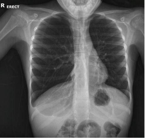
The patient, a 10-year-old child with a history of febrile syndrome, underwent urgent chest radiography, which revealed asymmetry between the hemithoraxes, with basal predominant hyperinflation and greater radiolucency in the right lung. A right paracardiac image suggestive of middle lobe atelectasis was noted, consistent with findings from previous studies dating back to at least 2015. A subsequent chest CT scan without intravenous contrast further delineated the extent of hyperinflation/ air trapping and pulmonary atelectasis in the right hemithorax, attributing these findings likely to a previous infectious process.
Discussion
The diagnostic imaging findings in this case are representative of the complexities associated with pediatric atelectasis. Radiography and CT scans serve as decisive tools for its diagnosis and monitoring. Atelectasis in children can result from various factors, including mucous plug formation, foreign body aspiration, and infections leading to airway obstruction [1]. The role of imaging, as highlighted in this case, extends beyond diagnosis to include guiding therapeutic interventions and assessing response to treatment.
State-of-the-art studies have increasingly emphasized the utility of advanced imaging techniques in pediatric atelectasis. For instance, several authors demonstrated the superior sensitivity of CT scans over traditional radiography in detecting subtle atelectatic changes and guiding surgical planning in pediatric populations [2]. However, X-ray tests - such as the one depicted in Figure 1 - are clear enough to guarantee an effective diagnosis. Furthermore, research by Hsu et al. [3] underscored the importance of integrating imaging findings with clinical assessment to optimize the management of atelectasis, particularly in identifying underlying causes and selecting appropriate interventions.
Conclusion
The present case underscores the critical role of imaging in diagnosing, managing, and understanding atelectasis in pediatric patients. Through a combination of chest radiography and CT scans, clinicians can gain comprehensive insights into the extent and nature of lung collapse, guiding effective treatment strategies. As the body of research grows, the integration of advanced imaging techniques with clinical practice promises to enhance the care of children with atelectasis, improving outcomes and quality of life.
References
- Johnston C, Carvalho WB de. Atelectasis: mechanisms, diagnosis and treatment in the pediatric patient. Rev Assoc Med Bras. 2008; 54: 455-60.
- Aquino SL, Schechter MS, Chiles C, Ablin DS, Chipps B, Webb WR. High-resolution inspiratory and expiratory CT in older children and adults with bronchopulmonary dysplasia. American Journal of Roentgenology. 1999; 173(4): 963-7.
- Hsu L, Green D, Chusid J, Talwar A, Shah R. Imaging of Atelectasis. Contemporary Diagnostic Radiology. 2013; 36(25): 1.


