1Department of Hepatobiliary Pancreatic Hernia Surgery, Guangdong Second Provincial General Hospital, School of Medicine, Jinan University, People’s Republic of China.
2Department of Anesthesiology Department, Guangdong Second Provincial General Hospital, School of Medicine, Jinan University, People’s Republic of China.
3Department of the First Surgical Ward of Minhang Campus, Guangdong Second Provincial General Hospital, People’s Republic of China.
4Department of Cardiothoracic Surgery, Guangdong Second Provincial General Hospital, People’s Republic of China.
5The Third Affiliated Hospital of Southern Medical University , People’s Republic of China.
6Department of Outpatient, Guangdong Second Provincial General Hospital, School of Medicine, Jinan University, People’s Republic of China.
#These authors have been equally contributed to this article.
Xiaofeng Li1; Cheng Li2; Xiancheng Zeng3
Email: gd2h_digitallab@163.com; 182838li@163.com; zxcq12333@163.com
Received : Dec 10, 2025 Accepted : Jan 05, 2026 Published : Jan 12, 2026 Archived : www.meddiscoveries.org
Biliary obstruction arises from various biliary diseases, and its alleviation can minimize obstructive jaundice and reduce risks of acute suppurative obstructive cholangitis. Bio-3D-printed biliary stents, building upon conventional designs, incorporate cells and drug-delivery capabilities to promote biliary repair and lower recurrence rates. This study provides a 20-year bibliometric analysis (2006–2025) to map global publication trends in bio-3D-printed biliary stents. Literature was retrieved from Web of Science on March 25, 2025. Using Cite Space, VOS viewer, and Microsoft Excel, we analyzed 169 relevant publications. The countries, institutions, journals, and authors with the highest number of publications were China (n=51), Zhejiang University (n=7), Scientific Reports (n=5), and van der Laan, Luc J.W.; Verstegen, Monique M.A.; Willemse, Jorke; Xiang, Yang (n=5), respectively. The most co-cited article was “Organ reengineering through development of a transplantable recellularized liver graft using decellularized liver matrix” from Nature Medicine (n=44). Keyword analysis identified research hotspots: hepatic tissue engineering, liver graft, 3D bioprinting, and biliary stent design. This bibliometric research comprehensively analyzes the field’s landscape, highlighting current trends and thematic focuses. These findings may guide clinicians and researchers toward future directions in bio-3D-printed biliary stent development.
Keywords: 3D-bioprinted; Biliary stent; Hepatic tissue engineering; Biliary obstruction; Bioengineering.
Biliary obstruction frequently arises from various benign and malignant biliary diseases, with the most common causes including: iatrogenic bile duct injury (primarily post-cholecystectomy), primary sclerosing cholangitis, post-liver transplantation biliary obstruction, common bile duct stenosis due to chronic pancreatitis, ampullary tumors, hilar cholangiocarcinoma, and pancreatic neoplasms [9,31]. Therapeutic approaches for biliary obstruction encompass percutaneous external drainage, endoscopic therapy, and surgical intervention. Endoscopic biliary stent implantation represents the most extensively utilized procedure [22,33], which involves deploying stents at stenotic sites to alleviate obstruction and restore biliary drainage [31]. Stent materials constitute a current research focus in this field, with emerging options including Plastic Stents (PSs), Self-Expandable Metal Stents (SEMS), uncovered SEMS (USEMS), and Polyurethane-Covered Stents (CSEMS) [13,14,35,36,42,43]. However, such commercially available stents typically lack personalized configurations, adaptability, and developmental potential [5,11,41].
Three-Dimensional (3D) printing technology provides a viable solution to these limitations. Since its conceptualization in the early 1980s, 3D printing has been widely applied in surgical planning, implant/prosthesis fabrication, and medical education [8,24]. Implant research within this domain includes biliary stent development. Notably, bioprinting—an advanced 3D printing technique utilizing “bio-inks” (cell-laden biomaterial composites)—enables fabrication of geometrically complex 3D tissue-mimicking constructs. This approach significantly enhances personalized biliary stents’ capabilities for drug delivery, tissue regeneration, and anti-obstruction efficacy, demonstrating substantial research value [27,28,32,47].
Bibliometrics employs mathematical-statistical methods to conduct qualitative-quantitative analysis of disciplinary literature. By evaluating publication impact and distribution patterns, this methodology identifies research trajectories and developmental trends [7], having been implemented in hepatobiliary, immunological, and oncological research [15,37]. Nevertheless, systematic analyses specifically addressing 3D-bioprinted biliary stents remain scarce. This study therefore applies bibliometric tools to comprehensively evaluate current research status and emerging directions in 3D-bioprinted biliary stent technology.
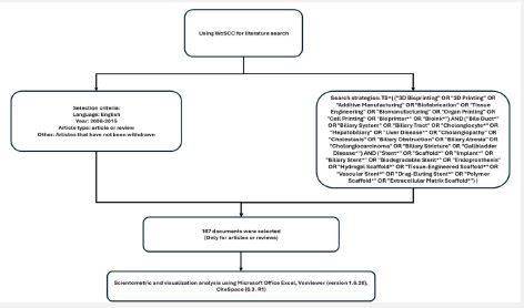
Data source and search strategy
The Web of Science Core Collection (WoSCC) database was selected as the data source for this study. The search strategy employed the following query: TS=((“3D Bioprinting” OR “3D Printing” OR “Additive Manufacturing” OR “Biofabrication” OR “Tissue Engineering” OR “Biomanufacturing” OR “Organ Printing” OR “Cell Printing” OR “Bioprinter*” OR “Bioink*”) AND (“Bile Duct*” OR “Biliary System” OR “Biliary Tract” OR “Cholangiocyte*” OR “Hepatobiliary” OR “Liver Disease*” OR “Cholangiopathy” OR “Cholestasis” OR “Biliary Obstruction” OR “Biliary Atresia” OR “Cholangiocarcinoma” OR “Biliary Stricture” OR “Gallbladder Disease*”) AND (“Stent*” OR “Scaffold*” OR “Implant*” OR “Biliary Stent*” OR “Biodegradable Stent*” OR “Endoprosthesis” OR “Hydrogel Scaffold*” OR “Tissue-Engineered Scaffold*” OR “Vascular Stent*” OR “Drug-Eluting Stent*” OR “Polymer Scaffold*” OR “Extracellular Matrix Scaffold*”)). The search was restricted to original research articles and review papers published in English between 1 January 2006 and 31 March 2025, with the Science Citation Index Expanded (SCIE) designated as the sole citation index. Literature retrieval and data download were completed on 25 March 2025 to eliminate bias from dynamic database updates.
Data extraction and analysis
The initial database search yielded 176 publications. Following the exclusion of 5 articles published prior to 2006, the remaining records were imported into Microsoft Office Excel 365. Subsequent manual screening identified and removed 2 retracted publications, resulting in a final corpus of 169 articles. Key bibliometric parameters—including country of origin, authorship, publication year, institutional affiliation, journal title, citation counts, and keywords—were systematically extracted. It should be noted that potential analytical discrepancies may arise due to inconsistent author abbreviation conventions and variations in journal citation styles. To mitigate retrieval bias, two investigators independently conducted the search procedures, with results demonstrating high inter-rater concordance.
Data visualization
Following data acquisition, all records were imported into VOS viewer (version 1.6.20) for comprehensive bibliometric analysis. This software enabled systematic examination of countries, authors, institutions, journals, citations, keywords, co-cited journals, and co-authorship networks. Within the VOS viewer-generated maps, distinct nodes represent individual elements of analysis, where node size corresponds to occurrence frequency or co-occurrence count, while connecting lines denote co-occurrence relationships. For enhanced visualization of international collaboration patterns, country-level publication relationships were further analyzed using SCImago. Subsequently, the dataset was processed through CiteSpace (version 6.3.R1) to leverage its specialized capabilities in keyword clustering, keyword frequency ranking, and timeline analysis, facilitating identification of research frontiers and emerging trends. All tabular data management and supplementary visualizations were performed using Microsoft Office Excel 365 to ensure comprehensive data organization and presentation.
The annual trend of publication
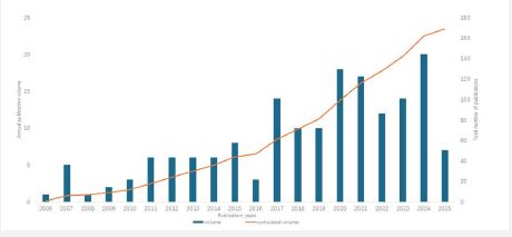
Our systematic review identified 167 publications related to 3D bioprinted biliary stents in the WoS database. As illustrated in (Figure 2), the annual publication count has demonstrated an overall upward trajectory since 2006. During the initial 11-year period (2006-2016), the field maintained relatively low output, with fewer than 6 publications per year. A notable inflection point occurred in 2017, when annual output surged to 14 articles. This increased productivity persisted throughout the subsequent 9-year period, consistently exceeding 10 publications annually. The peak output was observed in 2024 with 20 publications. Importantly, while the publication trend shows steady growth, the absolute numbers remain modest, indicating substantial untapped research potential in this emerging field.
Contributions of countries/regions
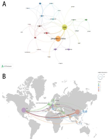
Our analysis identified 22 countries/regions contributing to this research field. The top three productive nations were China (n=51), the United States (n=49), and Japan (n=13), demonstrating their pivotal roles in 3D bioprinted biliary stent research. Network visualization revealed China and the United States as central hubs, evidenced by their largest node sizes and most extensive co-authorship links (indicated by thicker connecting lines). The United States exhibited particularly strong collaborative ties, with a total link strength of 31, reflecting both direct and indirect partnerships with numerous countries. While China’s overall link strength (n=17) was approximately half that of the United States, its primary collaborations involved other highly active nations (publications >10) including the United States, Germany, Iran, Japan, and South Korea.
Notably, despite their substantially lower publication outputs compared to China and the United States, the United Kingdom (n=15) and Sweden (n=14) demonstrated comparable collaborative influence, with link strengths nearly equivalent to China’s. (Figure 3) presents our VOS viewer and Scimago-generated visualization of these international research networks.
As detailed in (Table 1), while China marginally surpassed the United States in total publication count, citation analysis revealed a different pattern. The United States led in research impact with 1,863 citations - exceeding the combined total of China (1,079 citations) and Japan (749 citations), suggesting greater influence per publication despite slightly lower output volume.
Analysis of institutions
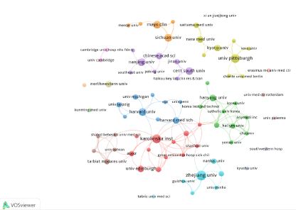
Our analysis identified 379 institutions that have published research on 3D bioprinted biliary stents. As shown in (Figure 4), among the 76 institutions with more than 2 publications worldwide, we highlight the top 10 most productive institutions (representing 15 organizations) for their outstanding contributions (Table 2). These leading institutions are distributed across five countries/regions: China, the United States, Japan, the United Kingdom, and Sweden.
Chinese institutions dominated the list, with five representatives. Zhejiang University emerged as the most productive, publishing 7 articles, followed by Central South University, Sichuan University, and Nanjing University with 5 publications each, while the Chinese Academy of Sciences contributed 4 articles. Sharing the second position were Sweden’s Karolinska Institutet and the University of Pittsburgh (USA), both with 6 publications. The remaining eight institutions from the US, Japan, and UK published between 4-5 articles each.
The network analysis revealed distinct collaborative clusters. The largest consortium centers around Karolinska Institute and Zhejiang University, forming an extensive international network spanning Europe, America, and Asia. A second notable cluster focused on Nanjing University and Central South University, though this collaboration appears primarily limited to China-US partnerships.
When examining collaboration strength, Karolinska Institute demonstrated the strongest ties (n=14), followed by Harvard Medical School (n=9). Interestingly, our analysis revealed a dissociation between publication quantity, collaborative networks, and research impact. Kyoto University, despite having the fewest publications (n=4), achieved the highest citation count (n=480), suggesting exceptional research quality or influence within this specialized field.
Related journals and co cited journals
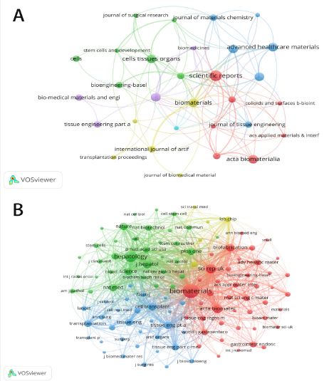
Our bibliometric analysis revealed that the 167 publications were disseminated across 108 academic journals (Figure 5). Among these, we identified 33 journals that published more than 2 articles on 3D bioprinted biliary stents. The most prolific journal was Scientific Reports (n=5, IF=4.6, JCR Q2), which not only showed the largest node in our network visualization but also demonstrated the densest connections, indicating both high productivity and significant collaborative influence. Following closely was the UK-based Biomedical Materials (n=4, IF=4.0, JCR Q3). Notably, among the top 10 most productive journals, Bioactive Materials from China boasted the highest impact factor, with 6 journals ranked in JCR Q1 or Q2 categories.
The co-citation analysis of 1,945 journals highlighted the 10 most frequently co-cited sources (Table 3), predominantly from the UK, USA, and Netherlands. Biomaterials emerged as the most influential with 580 co-citations, represented by the largest node in the red cluster of our visualization. Other highly co-cited journals included Hepatology (n=339) and Scientific Reports (n=235), both originating from developed nations. Interestingly, Nature Medicine (highest IF among analyzed journals) and its sister journal Nature showed nearly identical co-citation frequencies (147 each).
Analysis of authors and co-cited authors
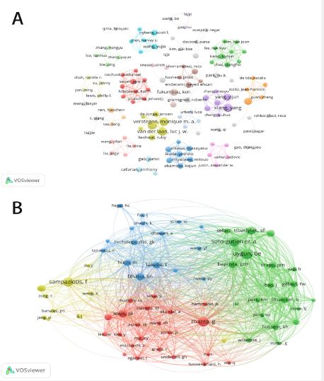
Our analysis identified 1,145 authors contributing to this research domain. (Table 4) presents the top 10 most productive researchers, with four scholars - van der Laan LJW, Verstegen MMA, Willemse J, and Xiang Y-tied for first position, each having published 5 articles. The remaining six authors in the ranking have all contributed more than 3 publications each.
The co-authorship network (Figure 6), constructed using authors with ≥2 publications, reveals distinct collaboration patterns. The four largest nodes correspond precisely to the most productive authors mentioned above. The visualization clearly demonstrates that most researchers primarily collaborate within their established teams, with limited cross-disciplinary cooperation. Notably, the top three authors - van der Laan LJW, Verstegen MMA, and Willemse J - form a tightly connected research cluster, indicating their affiliation with the same research institution or consortium.
Analysis of co-cited references and citation burstiness of references
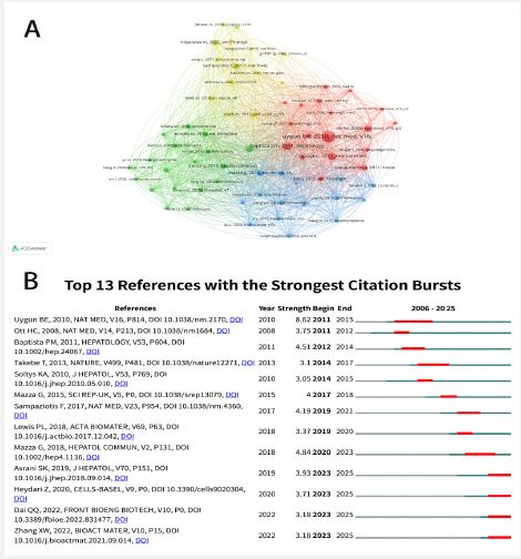
From the 169 publications, we selected 66 references with co-citation frequencies exceeding 5 to construct a co-citation network (Figure 7). (Table 5) lists the top 10 co-cited references, each receiving at least 16 co-citations. The most frequently co-cited work was Uygun BE’s 2010 Nature Medicine article “Organ reengineering through development of a transplantable recellularized liver graft using decellularized liver matrix” [44], with 44 co-citations. Ranking second was Baptista PM’s 2011 study “The use of whole organ decellularization for the generation of a vascularized liver organoid” [2] published in Hepatology (a leading hepatology journal), demonstrating 27 co-citations. Crapo PM et al.’s review “An overview of tissue and whole organ decellularization processes” [10] placed third with 25 co-citations. Notably, these three foundational references exhibit strong intellectual connections within the co-citation network, evidenced by their close nodal proximity and dense interconnecting lines, highlighting their pivotal contributions to 3D-bioprinted biliary stent research.
Using CiteSpace, we identified 13 publications with significant citation bursts (Figure 7B). In this visualization, each red or blue bar represents a one-year interval, with red bars indicating periods of citation bursts. The 13 publications exhibiting the strongest bursts span 2011-2025. Uygun BE’s landmark article demonstrated the most pronounced citation burst (intensity 8.62) during 2011-2015. Mazza G et al.’s Hepatology Communications paper “Liver tissue engineering: From implantable tissue to whole organ engineering” [30] ranked second, showing a citation burst (intensity 4.84) between 2020-2023. Subsequently, Baptista PM’s second-ranked co-cited reference displayed a citation burst (intensity 4.51) during 2012-2014.
Analysis of keywords
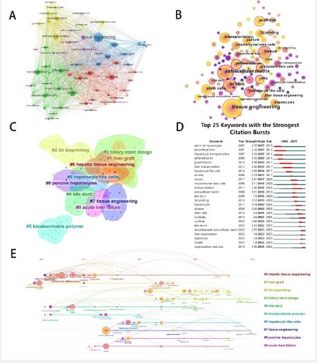
Keyword analysis identified 69 terms occurring ≥5 times in the literature, visualized using VOS viewer (Figure 8A). Each node’s size corresponds proportionally to the frequency of its corresponding keyword in the publications. “Tissue engineering” occupied the central position as the largest node, while other prominent nodes—including “extracellular matrix,” “tissue,” “in vitro,” “3D printing,” and “transplantation”—represented core research themes. CiteSpace-based keyword analysis (Figure 8B) yielded consistent thematic focus. (Figure 8C) displays the top 25 keywords exhibiting strongest citation bursts, with “adult rat hepatocytes” showing the earliest burst (2007-2010; strength=2.55) and “3d printing” demonstrating the most intense burst (2019-2020; strength=3.53). The keyword “mesenchymal stem cells” maintained sustained influence since 2008, peaking during 2018-2020 (strength=3.11).
CiteSpace clustering organized keywords into nine thematic groups (Table 6, Figure 8E), where the five largest clusters by keyword density were “hepatic tissue engineering” (n=21 keywords), “liver graft” (n=18), “3d bioprinting” (n=18), “biliary stent design” (n=15), and “bile duct” (n=8). Temporal evolution mapping (Figure 8D) identified “hepatic tissue engineering” as the longest-continuously researched cluster (2007-2023), contrasting with the short-lived “bioabsorbable polymer” cluster (2007-2015). Notably, “biliary stent design” (active since 2017) and “tissue engineering” remain dynamically investigated as of 2025.
| Countries/Regions | Documents | Citations | Total link strength |
|---|---|---|---|
| Peoples R China | 51 | 1014 | 17 |
| USA | 49 | 1863 | 31 |
| Japan | 13 | 749 | 6 |
| Germany | 13 | 401 | 8 |
| Iran | 11 | 223 | 7 |
| South Korea | 11 | 237 | 4 |
| England | 10 | 146 | 15 |
| Italy | 9 | 189 | 9 |
| Sweden | 9 | 113 | 14 |
| India | 9 | 188 | 4 |
| Zhejiang University (China) | 7 | 291 | 8 |
| Karolinska Institute (Sweden) | 6 | 63 | 14 |
| University of Pittsburgh (United states) | 6 | 458 | 7 |
| Central South University (China) | 5 | 58 | 4 |
| Harvard University (United states) | 5 | 267 | 7 |
| Mayo clinic (United states) | 5 | 75 | 5 |
| Nanjing University (China) | 5 | 173 | 5 |
| Sichuan University (China) | 5 | 48 | 5 |
| Chinese Academy of Sciences (China) | 4 | 83 | 5 |
| Hanyang University (South korea) | 4 | 141 | 8 |
| Harvard Medical School (United states) | 4 | 151 | 9 |
| Kyoto University (Japan) | 4 | 480 | 4 |
| Nara Medical University (Japan) | 4 | 463 | 5 |
| Northwestern University (United states) | 4 | 147 | 0 |
| The University of Edinburgh (England) | 4 | 28 | 3 |
| Source | Documents | IF2024 | JCR2024 | Source | citations | IF2024 | JCR2024 |
|---|---|---|---|---|---|---|---|
| Scientific Reports (England) | 5 | 4.6 | Q2 | Biomaterials (Netherlands) | 580 | 14.0 | Q1 |
| Acta Biomaterialia (Netherlands) | 4 | 9.7 | Q1 | Hepatology (United states) | 339 | 13.5 | Q1 |
| Advanced Healthcare Materials (Germany) | 4 | 10.0 | Q1 | Scientific Reports (England) | 235 | 4.6 | Q2 |
| Biomaterials (Netherlands) | 4 | 14.0 | Q1 | Acta Biomaterialia (Netherlands) | 198 | 9.7 | Q1 |
| Biomedical Materials (England) | 4 | 4.0 | Q3 | Journal of Hepatology (Netherlands) | 178 | 25.7 | Q1 |
| Cells Tissues Organs (Switzerland) | 4 | 2.5 | Q4 | Biofabrication (England) | 157 | 9.1 | Q1 |
| Frontiers in bioengineering and Biotechnology (Switzerland) | 4 | 5.7 | Q2 | Cell Transplantation (United states) | 155 | 3.1 | Q3 |
| Tissue engineering part c-methods (United states) | 4 | 3.5 | Q3 | Nature Medicine (England) | 147 | 82.9 | Q1 |
| Bio-medical materials and engineering (Netherlands) | 3 | 2.0 | Q4 | Tissue Engineering Part A (United states) | 147 | 3.5 | Q3 |
| Bioactive materials (China) | 3 | 18.9 | Q1 | Nature (England) | 138 | 64.8 | Q1 |
| Author | Documents | Co-cited author | Co-citations |
|---|---|---|---|
| Van der laan, luc j. W. | 5 | Uygun, be | 52 |
| Verstegen, monique m. A. | 5 | Mazza, g | 46 |
| Willemse, jorke | 5 | Sampaziotis, f | 43 |
| Xiang, yang | 5 | Soto-gutierrez, a | 43 |
| Fukumitsu, ken | 4 | Badylak, sf | 36 |
| Yang, yijun | 4 | Bhatia, sn | 33 |
| Zhang, lei | 4 | Lewis, pl | 32 |
| Aikawa, masayasu | 3 | Baptista, pm | 30 |
| Callanan, anthony | 3 | Takebe, t | 30 |
| Choi, dongho | 3 | Ott, hc | 29 |
| Cited reference | Citations |
|---|---|
| Uygun Be, 2010, Nat Med, V16, P814 | 44 |
| Baptista Pm, 2011, Hepatology, V53, P604 | 27 |
| Crapo Pm, 2011, Biomaterials, V32, P3233 | 25 |
| Ott Hc, 2008, Nat Med, V14, P213 | 21 |
| Mazza G, 2015, Sci Rep-Uk, V5 | 19 |
| Sampaziotis F, 2017, Nat Med, V23, P954 | 19 |
| Takebe T, 2013, Nature, V499, P481 | 19 |
| Mazza G, 2018, Hepatol Commun, V2, P131 | 16 |
| Miyazawa M, 2005, Am J Transplant, V5, P1541 | 16 |
| Soto-Gutierrez A, 2011, Tissue Eng Part C-Me, V17, P677 | 16 |
| Cluster ID | Size | Silhouette | Mean (Year) | Label (LSI) | Label (LLR) | Label (MI) |
|---|---|---|---|---|---|---|
| 0 | 21 | 0.969 |
Tissue engineering; Extracellular matrix; Mesenchymal stromal cells; Cholangiocarcinoma; Physiological microenvironments; Transplantation; Expression; Cell derived hepatocytes; Liver; Failure |
Hepatic tissue engineering (10.67, 0.005); Bioreactor (7.35, 0.01); Expression (7.09, 0.01); Bile duct (7.07, 0.01); Decellularization (7.09, 0.01) |
Liver-as-chip (0.63); Pancreatitis (0.63); pH |
|
| 1 | 18 | 0.821 |
Tissue engineering; Liver graft; Extracellular matrix; Decellular liver; Natural scaffold; Differentiation; Rheological characterization; Lung cholangiocytes; Scaffold |
Liver graft (8.39, 0.005); Rheological characterization (4.18, 0.05); Functional human (4.18, 0.05); Submucosal bone (4.18, 0.05); Matrix (4.18, 0.05) |
Rheological characterization (0.42); Functional human |
|
| 2 | 18 | 0.843 | 2021 |
Tissue engineering; 3d bioprinting; Hepatocyte; 3d bioprinted liver; Hepatic cell; Electronic 3d printing; Cell-derived; Patient-specific bile duct; Hepatocyte; Tissue engineering |
3d bioprinting (5.39, 0.05); Disease (4.61, 0.05); Artificial multi-cellular tissue (4.61, 0.05); Hepatoblast-like cell (4.61, 0.05); 3d printing template (4.61, 0.05) |
Disease (0.33); Artificial multi-cellular tissue |
| 3 | 15 | 0.861 | 2020 |
Tissue engineering; Artificial bile duct; Nano-fabrication coating; Localized tissue construct; Malignant biliary obstruction; Biliary stricture; Biliary tract disease; Therapeutic intervention |
Biliary stent design (5.28, 0.05); Bile duct disruption (5.28, 0.05); Tissue-engineered (5.28, 0.05); TEG (5.28, 0.05); 3d bioprint (5.28, 0.05) |
Biliary stent design (0.2); Bile duct disruption |
| 4 | 14 | 0.597 | 2019 |
3d printing; Magnetic resonance imaging; Partial hepatectomy; Bump graft; Liver tissue; Engineering; Hepatobiliary transplantation; Humanized |
Bile duct (26.14, 0.01); Magnetic resonance imaging (mri) (10.32, 0.005); 3d printing (7.64, 0.01); Hepatocyte transplantation (6.65, 0.01); Gelatin/methacryloyl (Gelma) (5.14, 0.05) |
Gelatin/methacryloyl (gelma) (0.32); Whole-organs |
| 5 | 12 | 0.856 | 2018 |
Biodegradable polymer; Artificial bile duct; Tissue engineering; Bile duct stenosis; Extracellular matrix; Regeneration tissue engineering; Engineering bile duct; Biliary tract disease; Basic biofabrication; Bile duct stenosis |
Bioabsorbable polymer (15.11, 0.04); Artificial bile duct (15.51, 0.01); Bile duct stenosis (7.55, 0.05); Collagen (7.55, 0.01); Basic biofabrication factor (7.55, 0.01) |
Bile duct stenosis (0.5); Collagen (0.5) |
| 6 | 11 | 0.332 | 2015 |
Liver tissue engineering: 3D bioprinting; Cell culture; Physiological relevance; Mesenchymal stem cell; Proliferation; Hepatic differentiation; Hepatocyte function; Regenerative tissue |
Hepatocyte-like cells (6.2, 0.005); Liver tissue engineering (4.7, 0.005); Tissue engineering (4.7, 0.005) |
Hepatocyte-like cells (0.26); Hepatic tissue (0.26) |
| 7 | 10 | 0.718 | 2018 |
Tissue engineering; Common bile duct; Extracellular matrix; Vascularization strategy; 3D bioprinting; Liver regeneration; Decellularization; Liver scaffold; Liver niche |
Tissue engineering (14.1, 0.001); Liver tissue engineering (5.1, 0.05); Regenerative medicine (4.2, 0.05); Tissue repair (3.5, 0.1); Decellularized liver (3.1, 0.1) |
Vascularization strategies (0.36); In vitro and in vivo |
| 8 | 9 | 0.952 | 2016 |
Regenerative medicine; Tissue engineering; Organ engineering; Liver transplantation alternatives; Tissue engineering; Tissue modeling; Bioengineers; Tissue engineering; Organ engineering |
Primary hepatocytes (10.2, 0.005); Stem cells (7.8, 0.01); Regeneration (5.1, 0.05); Tissue engineering (4.7, 0.005); Working scaffold (4.2, 0.05) |
Regeneration (0.23); Tissue engineering; Modalities |
| 9 | 6 | 0.954 | 2015 |
Liver tissue engineering; Angiogenesis; Hepatic differentiation; Human-induced pluripotent stem cell; iPSC; Vascular mesenchymal stem cell; Vasculogenesis; Animal model; Hypoxia; Proliferation |
Acute liver failure (5.5, 0.05); Whole liver (3.9, 0.05); Spheroids (3.9, 0.05); Liver regeneration (3.9, 0.05); Hypoalbuminemia (3.9, 0.05) |
Whole liver (0.19); Spheroids (0.19) |
Annual publication trend analysis
Over the past two decades, 379 institutions across 22 countries/regions have published 167 articles on 3D-bioprinted biliary stents in 108 academic journals. As depicted in (Figure 2), the publication volume demonstrates a marked upward trajectory, signifying the growing significance of 3D-bioprinted biliary stents within related research domains. This trend is particularly pronounced during the 2017-2025 period. The pre-2017 research landscape was characterized by modest growth, with annual publications consistently below 6 articles. In contrast, the 2017-2025 interval witnessed a substantial expansion in output. Notably, the current year (2025) has already yielded 7 publications as of March—reinforcing sustained scholarly interest among specialists in this evolving field.
Geographical distribution and research contributions
Among the top 10 most productive countries/regions, developed nations dominate the research landscape, with only three developing economies represented: China, India, and Iran. This distribution underscores significant disparities in research capacity between developed and developing regions. Developing nations encounter persistent challenges in advancing 3D bioprinting research—particularly regarding funding constraints, limited access to cutting-edge scientific resources, and insufficient technological infrastructure. Nevertheless, these regions actively engage in knowledge transfer from developed countries, with China demonstrating exceptional progress. All contributing nations strive to advance this emerging field through substantive scholarly contributions.
Geographically, these leading countries/regions span three major areas: Europe, Asia, and North America, collectively maintaining rigorous research standards. The United States not only leads in research output but has also established extensive collaborative networks with European partners including Italy, Germany, and France. Such international cooperation has accelerated the application of 3D bioprinting in biliary stent development and propelled technological innovation. Within Asia, China distinguishes itself through substantial research output, with prolific publication activity in recent years.
Notably, while the United States trails China marginally in publication volume, it demonstrates significant dominance in citation impact. This divergence suggests U.S. research contributions generate greater scholarly influence per publication compared to other nations. The observed geographical patterns reflect both existing research infrastructure advantages and emerging global knowledge-exchange dynamics that continue to shape the field’s evolution.
Institutional productivity analysis
The analysis of the top 10 most productive institutions reveals a clear dominance by Chinese and American research centers. As illustrated in (Figure 4), among the top 10 institutions with the highest output (a total of 15), China and the United States each claim 5 representatives, with the remaining 5 institutions originating from Japan, South Korea, the United Kingdom, and Sweden. These organizations have made substantial contributions to advancing the application of 3D bioprinting in biliary stent development.
Zhejiang University (China) emerges as the most prolific institution with 7 publications. It is closely followed by Sweden’s Karolinska Institutet and the University of Pittsburgh (USA), underscoring their significant influence in the field. Notably, while the University of Pittsburgh ranks among the top three in publication output, it demonstrates exceptional citation performance. Similarly noteworthy is Japan’s Kyoto University, which despite having fewer publications, achieves the highest citation count (n=480) - a testament to the profound academic impact and research excellence of these institutions.
The extensive collaborative network centered around Karolinska Institutet and Zhejiang University demonstrates particularly strong academic synergy. This robust institutional framework highlights the exceptional research capabilities and innovative potential of investigators working within these centers on 3D bioprinted biliary stent development. The observed patterns suggest that while publication quantity reflects research activity, citation impact better indicates scholarly influence and technological advancement in this specialized field.
Journal and citation landscape analysis
Our journal analysis reveals that Scientific Reports leads in publication output within this field. As a Q2 journal with an Impact Factor (IF) of 4.6, it also ranks third in citation frequency (n=235). Notably, Tab. 3 demonstrates that both the most productive and most cited journals predominantly originate from Europe, including high-impact titles such as Nature (IF>25), Nature Medicine (IF>25), and Journal of Hepatology (IF>25). This European dominance underscores the region’s pivotal role in advancing 3D bioprinting technologies for biliary stent applications, where researchers consistently publish high-quality studies that shape clinical adoption of these innovations. We recommend that investigators prioritize engaging with these high-impact journals—both as sources of cutting-edge knowledge and as platforms for disseminating their own findings to maximize scholarly influence.
In terms of citation impact, the most cited work is the seminal Nature Medicine paper, “Organ reengineering through development of a transplantable decellularized liver graft using decellularized liver matrix” [44]. This article also exhibited the strongest citation burst, coinciding precisely with the period of accelerated growth in publication volume for this field. The study introduced a breakthrough technique for effective recellularization of liver grafts in vitro, maintaining cell viability and function—an advancement that laid the foundation for modern tissue engineering approaches and directly enabled subsequent progress in 3D-bioprinted biliary stents.
Equally significant is the second-ranked citation burst article, “Liver tissue engineering: From implantable tissue to whole organ engineering” (2018), which rapidly gained widespread recognition. This work highlighted the previously understudied biliary system within liver tissue engineering, marking a paradigm shift that redirected bioprinting research toward addressing biliary applications—a transition of profound clinical relevance.
Keyword analysis and research hotspots
Through co-occurrence analysis of 69 keywords derived from 169 articles, we gained valuable insights into current research directions and emerging hotspots within the field. As illustrated in (Figure 8), the majority of studies cluster around themes such as “Liver Tissue Engineering,” “3D Bioprinting,” and “Biliary Stent Fabrication.”3D bioprinting has advanced significantly in recent years, evolving from the printing of simple biomaterials to the printing of living cells. Living cell 3D printing utilizes cells and biomaterials as “bio-inks,” enabling precise spatial control over cell placement and the surrounding microenvironment. This maximizes the simulation of in vivo conditions for constructing in vitro tissue or organ models, demonstrating immense potential in organ reconstruction, drug screening, and mechanistic studies [40].
As revealed by keyword clustering and co-occurrence, within hepatobiliary research, 3D bioprinting was first applied to liver tissue engineering. Initial studies employed Two-Dimensional (2D) cell culture on dishes, where cells grew in monolayers. This approach lacks the complex cell-cell and cell-matrix interactions found in vivo. In contrast, cells grown under Three-Dimensional (3D) conditions exhibit advantages in cellular organization and the expression of cell-specific genes, more closely resembling in vivo biological behavior. Consequently, many 3D printed culture systems are now recognized as more accurate predictors of cellular responses than 2D cultures [39]. Following this rationale, researchers have utilized bioprinted liver scaffolds to replicate the complexity of tissue architecture. This approach holds promise for overcoming limitations in current cell therapy strategies, and the development of implantable engineered liver tissue offers significant potential for treating liver failure-related diseases [1,2]. Zhong et al. [48] constructed hydrogel scaffolds incorporating the human normal hepatocyte cell line HL-7702. Transplantation of these scaffolds into liver-injured nude mice resulted in superior liver function compared to the control group [48]. Kang et al. [16] developed a 3D liver tissue model using mouse embryonic fibroblast-derived induced hepatocytes (miHeps). Expression of liver-specific markers (albumin, ASGR1, HNF4α) progressively increased during in vitro culture. Transplantation of these constructs into liver-injured mice led to enhanced cell proliferation and higher albumin expression [16]. These studies collectively demonstrate the considerable therapeutic potential of 3D bioprinted liver scaffolds for liver regeneration. The bile ducts are integral components of liver tissue, containing cholangiocytes (biliary epithelial cells). Advances in liver tissue engineering and the successful development of viable liver tissue scaffolds suggest that bioprinted biliary scaffolds for repair functions possess analogous potential.
Bile duct injury is a severe complication of biliary surgery [17]. Patients suffering such injuries face risks including bile leakage, biliary obstruction, and even liver failure, with biliary obstruction during repair being the core challenge. Treatment for biliary obstruction has evolved significantly, progressing from traditional open surgery to percutaneous external drainage and, more recently, endoscopic techniques. Endoscopic therapy, with its advantages of minimal invasiveness, speed, and precision, has become paramount for the diagnosis and treatment of biliary diseases. Endoscopic biliary stent placement is now the most widely used procedure for managing biliary obstruction [4,20]. While conventional stents are effective for benign biliary obstruction, they often provide only symptomatic relief for injuries requiring ductal repair and can be associated with significant complications [6]. Research indicates that bile ducts can also be engineered. Baptista et al. [2,24] pioneered this area, demonstrating that decellularized liver matrices (scaffolds retaining only the extracellular matrix) could support the differentiation of fetal hepatoblasts into both cholangiocyte and hepatocyte lineages. Building on this, Lewis et al. further showed that murine cholangiocytes and human Huh7 cells could form liver tissue incorporating a common bile duct structure [23]. This implies that incorporating such cells into bioprinted biliary scaffolds could make the repair of bile duct injuries via stents a realistic prospect.
As shown in (Figure 8), research activity involving the keyword “3D Bioprinting” surged after 2016, leading to a corresponding increase in its application for biliary stents. Traditionally implanted biliary stents were fabricated from plastic, metal, or other polymeric materials. However, such stents carry a significant risk of complications including cholecystitis, pancreatitis, restenosis, erosion/embedding into the duct wall, and biliary perforation, causing additional patient harm [3,18,19].
In contrast, 3D bioprinted biliary stents offer excellent biocompatibility, stable mechanical properties, and biodegradability, making them ideal for supporting and repairing damaged bile ducts. This technology involves the layer-by-layer deposition of biomaterials, cells, and biomolecules to create 3D biological constructs. Primary printing modalities include extrusion-based printing, inkjet/droplet printing, and Digital Light Processing (DLP) [29]. A key advantage of 3D bioprinting is its capacity to create patient-specific structures tailored to precise anatomical and physiological requirements. Although the process can be time-intensive, it allows for the precise placement of cells and biomaterials, facilitating the incorporation of multiple cell types and materials to form tissues with complex cellular and functional compositions.
Zong et al. developed a bilayered tissue-engineered common bile duct stent. The inner layer, composed of densely structured, slow-degrading Polycaprolactone (PCL), provided mechanical support. The outer layer, fabricated via 3D printing using loosely structured, fast-degrading Poly (Lactic-co-Glycolic Acid) (PLGA), promoted cell growth. Mesenchymal Stem Cells (MSCs) were seeded onto this bilayered structure using bioprinting techniques, and the scaffold demonstrated favorable therapeutic outcomes in vivo [49]. Other studies have focused on manufacturing absorbable 3D bioprinted biliary stents with functional properties, significantly advancing their clinical applicability [25,45].
Despite the promise, 3D bioprinted biliary stents face challenges. Certain biomaterials can provoke inflammatory and immune responses, leading to lymphocyte and fibroblast infiltration and proliferation within the bile duct wall [26,34,38]. Consequently, bioengineered biliary stents carry a potential risk of inducing hyperplasia [21]. However, ongoing research is actively addressing these issues, focusing on enhancing biocompatibility and mitigating hyperplasia risks [20,46].
Our study conducted a comprehensive visualization analysis of 169 articles published between 2006 and March 25, 2025, focusing on 3D bioprinted biliary stent research. As previously discussed, this field remains in its developmental stage, with the United States and China emerging as leading contributors. The primary research clusters concentrate on “hepatic tissue engineering,” “3D bioprinting,” and “biliary stent fabrication.”
Current advancements demonstrate that 3D-bioprinted biliary stents incorporating bioinks with mesenchymal stem cells or cholangiocytes exhibit superior performance characteristics, making them ideal candidates for supporting and repairing damaged bile ducts. However, significant challenges persist, including suboptimal fabrication success rates and the potential for biomaterial-induced cholangiocyte hyperplasia - issues that demand careful consideration and will likely dominate future research priorities.
While 3D-bioprinted biliary stents show considerable promise, substantial work remains before clinical translation can be realized. Our study provides a thorough overview of current research outputs in this field, offering valuable reference points for scholars investigating similar topics. The findings highlight both the remarkable progress achieved to date and the critical knowledge gaps that must be addressed to advance this promising technology toward clinical application.
Acknowledgements: This research was supported by Guangdong Medical Science and Technology Research Fund (no: A2024472), in part by 2024 Guangzhou Science and Technology City School Joint Project (no. 2024A03J0988).
Author contributions: Xiancheng Zeng, Cheng Li and Xiaofeng Li contributed to the conception and design of the study; Jiawei Qin, Ping Han, Lei Zhang, Wenjun Zhou, Renpeng Duan, Ruijian Liu performed the research and analyzed the data; Xiaofeng Li wrote the paper.
Conflict of interest: The authors declare that they have no conflict of interest.
Authorship contribution: Jiawei Qin: Formal analysis, Data curation, Writing – original draft. Ping Han: Formal analysis, Data curation. Lei Zhang: Formal analysis, Data curation. Wenjun Zhou: Formal analysis, Data curation. Renpeng Duan: Formal analysis, Data curation. Ruijian Liu: Formal analysis, Data curation. Xiancheng Zeng: Conceptualization, Methodology. Cheng Li: Conceptualization, Methodology. Xiaofeng Li: Conceptualization, Methodology.
