1Department of Pharmacology, Grace College of Pharmacy, Kodunthirapully, Palakkad 678004, Kerala, India.
2Affilated to Kerala University of Health Sciences, Thrissur 680596, Kerala, India.
Nishfar K
Tel: 8590117740;
Email: nishfar.k77@gmail.com
Received : Jan 08, 2025 Accepted : Feb 05, 2025 Published : Feb 12, 2025 Archived : www.meddiscoveries.org
Using an in-silico technique, the current work sought to find novel drug-like compounds with anticancer characteristics. The human topoisomerase 1-DNA complex (PDB ID 1A35) is selected as the protein. Ketoconazole, which is an antifungal agent is used as the ligand in this study along with irinotecan as the standard were sketched using chemdraw ultra 8.0 and converted to 3D forms. Both the protein and ligand were energy minimised for docking studies. Ligands are evaluated by Lipinski’s rule of five and docked with 1A35 enzyme with autodock vina in PyRx software. The resulting binding energy for ketoconazole is -8.4 kcal/mol and for irinotecan is -8.4 kcal/mol. The visualization of interactions is done using Biovia Discovery Studio 2024 Client. The study reveals the possibility of Ketoconazole in relevant anticancer activity, focusing on its drug-target interaction, contributes to anti-cancer drug discovery.
Keywords: Ketoconazole; Molecular docking; In silico study; AutoDock vina; Topoisomerase I; Lipinski’s rule of five; Anticancer agents.
Cancer is a group of linked disorder characterised by uncontrolled division of abnormal and non-productive cells which continue to exist and proliferate within the specific site of the body, may or may not spread to the surrounding tissues [1,2]. Cancer is a gene related condition which is caused by variety of genetic or habitual or environmental conditions such as UV or ionizing radiations, alcohol, tobacco smoke, aflatoxins, arsenic, or other chemicals or biological agents (virus or bacteria) [3,4,9]. Cancer cells are having multiple faulty chromosomes and DNA repair mechanism, which dodges apoptosis and proliferate continuously and exists as immature cells [13,15].
According to NCDIR India in 2022, about 1.4 lakhs of new cases were reported which makes probably one in nine people will face cancer in their lifetime [3]. Also, lung cancer is prominent in males and breast cancer in females. According to non-communicable diseases, cancer ranked second in death rate by 18.1% [11].
Since now, there is no ideal treatment protocol approved for cancer. Recent development of drugs in cancer treatment is either costly or are having numerous side effect including fatigue, nausea, vomiting, hair loss, anaemia, neutropenia, lymphedema, and secondary carcinoma [4,5].
This study is about in silico evaluation of ketoconazole as a topoisomerase I inhibitor in cancer treatment. Ketoconazole is currently marketed as an antifungal drug for topical administration [20]. It acts by inhibiting the fungal sterol synthesis, inhibiting the fungal cell wall and fungal proliferation [12]. It is expected to inhibit the cancer cells by topoisomerase I inhibition.
In this paper we are reporting the docking analysis of ketoconazole against topoisomerase I enzyme. The docking was performed to predict the binding affinity of the ketoconazole against this enzyme. Thus, the study produces useful data on repurposing of ketoconazole in treatment of cancer. The reference used is irinotecan.
In this study a number of softwares are used for different purposes.
| Sl no. | Software’s | Purposes |
|---|---|---|
| 1 | ChemDraw Ultra 8.0 | to create the 2D structure of ligands. |
| 2 | Chem3D Pro 8.0 | to create3D model and for ligand energy minimization |
| 3 | MOE (Molecular Operating Environment) | For protein energy minimization |
| 4 | PyRx | Autodock vina software virtual screening tool |
| 5 | Biovia Discovery Studio | Visualisation and Interpreting docking results |
Preparation of protein
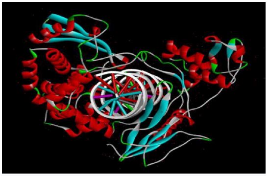
Human topoisomerase 1/DNA complex protein having PDB ID 1A35 was downloaded from www.pdb.org in PDB format. It was then energy minimised using MOE software.
Preparation of ligand (ketoconazole): The 2D structure of ketoconazole was prepared using CHEMDRAW ULTRA 8.0 software. It was then converted to 3D using chem 3D pro-8.0. also, the energy minimization was done with MOE software and was saved in PDB format.
Lipinski’s rule: Lipinski’s rule of five was used to predict the pharmacokinetic property of a drug based on it’s certain physiochemical parameters such as molecular weight (< 500 Da), number of hydrogen bond acceptors (< 10), number of hydrogen bond donors (< 5), partition coefficient ie; log P value (< 5). According to this rule, the ligand must not violate more than one condition [6].
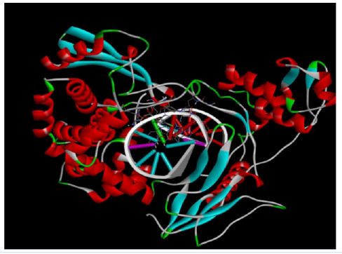
Docking studies: The ligand (ketoconazole) is then made to interact with the protein (1A35) and molecular docking is done. Docking studies provides with information regarding the interaction of the drug with the enzyme, binding affinity and binding energy. The molecular docking helps to simulate these interactions in detail.
In this study the docking was performed in pyRx software, by uploading the energy minimised protein (1A35) and ligand (ketoconazole) in PDB format, and performed the docking using a virtual screening tool, autodockvina pyRx. The docking results was analysed using BioVia discovery studio.
The docking results were compared with the docking of standard drug of irinotecan in the similar way as that of the molecular docking of ketoconazole. The different binding interactions and binding energy will be compared.
| Lipinski’s parameter | Values of ketoconazole | Limit |
|---|---|---|
| Molecular weight | 517.40Da | < 500Da |
| Number of hydrogen bond acceptors | 5 | < 10 |
| Number of hydrogen bond donors | 0 | <5 |
| Log P value | 3.42 | <5 |
| Number of violations | 1 |
Lipinski’s rule
The drug or ligand, ketoconazole passes the rule. Lipinski’s rule states four criteria, out of which the drug should not violate not more than one rule. The criteria include, the molecule should be within the limit of <500Da molecular weight, <10 hydrogen bond acceptors, <5 hydrogen bond donors, <5 log P value, for optimum pharmacokinetic property. Ketoconazole passes all the conditions except the molecular weight (517.40Da). However, the drug passes lipinski’s rule of five [6].
Docking studies
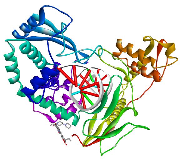
The binding energy of the ketoconazole was found to be -8.2 kcal/mol-1, while that of the standard drug (irinotecan) was found to be -8.4 kcal/mol-1. This shows that ketoconazole shows almost similar binding affinity when compared to standard. In molecular docking studies, binding energy describes the stability of the interaction [7,14]. The more negative the binding energy, more stronger the binding affinity, the more stable the ligand-receptor interaction [14]. The RMSD (root mean square deviation) value will be used to measure the closeness of the geometry of protein-ligand interaction to that of the predicted geometry [8]. In this study, the RMSD value is 0.
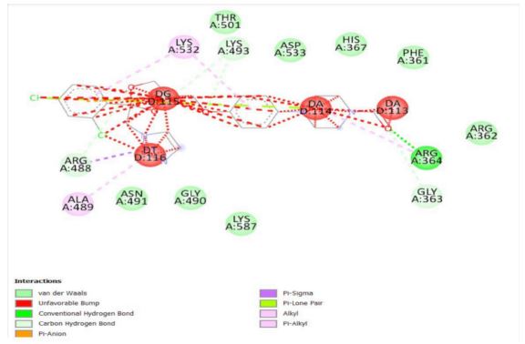
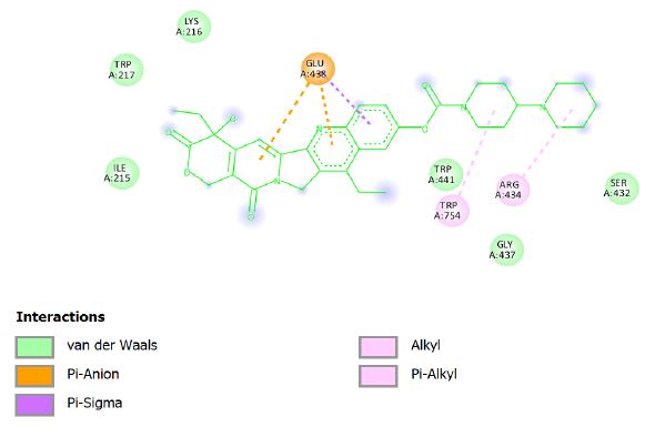
In contrast, the drug ketoconazole shows negative binding affinity values much alike that of irinotecan. The amino acid residue involved in the interactions of ketoconazole with enzymes are ARG A:364 by conventional hydrogen bond, LYS A:493, ARG A:488, GLY A:363 by Carbon-Hydrogen bond, ARG A:488 by Pi-sigma residue, DG D;115 by Pi-lone pair residue, LYS A:532, ALA A:489 by Pi-Alkyl residue, and THR A:501, ASP A:533, HIS A:367, PHE A:361, GLY A:363, LYS A:587, GLY A:490, ASN A:491 by van der Waals forces, which together contributes to the topoisomerase 1/DNA complex inhibitory actions and corresponding anti-cancer activity.
Cancer is a group of disease which is first reported even before 3000 BC [19] and the study of cancer and its treatment has been started by Hippocrates [3], till now an ideal treatment for cancer has not been developed [18]. In this study, the evaluation of the effect of ketoconazole on topoisomerase I enzyme was performed by molecular docking technique. It results that ketoconazole binding energy of -8.2 kcal/mol-1, which is closer to binding energy of -8.4 kcal/mol-1 by irinotecan (standard). Taking consideration of the difference in cost effectiveness, effectiveness, adverse reactions and resistance in different individuals, ketoconazole could be effectively used for anti-cancer property.
Acknowledgment: This work was supported by Kerala University of Health Sciences, Thrissur, Kerala Under UG Research Appreciation Award Scheme 2024.
